
How Artificial Intelligence Is Helping Doctors Detect Lung and Heart Disease
Published 4/1/26
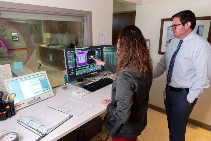 Radiologist Seth Kligerman, MD, pulls up a series of scans on his monitor — blobs of black and gray to the untrained eye. “These are CT scans of lungs. One thing we are trained to look for is nodules. While the vast majority of nodules we encounter are benign, some could potentially represent early lung cancer,” he says, noting that for each CT scan, radiologists review roughly between 400-600 slices, encompassing the entire volume of the lungs. Given how much visual data a radiologist needs to assess any scan, one can imagine that small nodules, many measuring less than 5 mm in size (about the width of a pencil), could be easily overlooked. “Even experienced radiologists who are subspecialty-trained in chest imaging can miss lung nodules,” says Dr. Kligerman. “And this is where AI-enhanced technology can be a big help.”
AI algorithms have the potential to improve diagnosis for diseases that affect different parts of the body, making them ideal for multispecialty hospitals. Dr. Kligerman moves on from CT scans of the lungs to CT images of the heart and coronary arteries, demonstrating how some algorithms can help doctors quantify the narrowing of the coronary arteries due to atherosclerotic disease, which can reduce the blood flow to the heart, leading to muscle damage. It also identifies subtypes of coronary plaques that are more commonly associated with an increased risk heart attack. He points to spots where the AI has marked areas of coronary artery atherosclerosis. “Here is an area of mild stenosis in one of the coronary arteries that I estimated to be a 42% stenosis and the algorithm estimated to be a 40% stenosis,” he says, making a note. “The readouts are very detailed. However, they are not always 100% accurate. We need to review each result closely, as any AI algorithm will make mistakes. For instance, this program may significantly underestimate or overestimate a stenosis. It is imperative that we review all output from AI algorithms ourselves before coming to a conclusion. It also helps that we have a brand new, state-of-the-art photon counting CT scanner that has a spatial resolution as low as 0.2 mm (about the width of 2 pieces of hair) that gives us clearer, more detailed images.”
Using AI to Detect Lung Disease Earlier
In addition to the commercially available AI-based tools being used by Dr. Kligerman and others, researchers at National Jewish Health are developing methods to evaluate diseases like emphysema and interstitial lung disease (ILD). “AI enables objective measurement of complex patterns like lung fibrosis (scarring) that are very difficult to quantify visually,” says Stephen Humphries, PhD, Director of the Quantitative Imaging Lab at National Jewish Health.
Dr. Humphries, whose background is in physics and biomedical engineering, has spent years developing and validating AI analysis models for lung images. “We’ve developed tools that can detect and quantify features in CT scans,” he explains. “These tools open up possibilities for clinical care and research. We’ve shown that automated analysis is more precise than visual assessment and can perceive clinically meaningful changes that are difficult for the human eye to detect. Increasingly, we’re also connecting scanning results with genetic information or blood-based biomarkers to better understand the causes and progression of ILD.”
This combination of different technologies may also enable doctors to detect disease earlier and track its progression with greater accuracy. “The amount of fibrosis, or scarring, in the lung — what percentage of the lung appears to be affected — is a strong indicator for disease,” Dr. Humphries says. “This can help inform treatment decisions. We can also detect small amounts of lung fibrosis in people at risk of developing ILD.” The AI system Dr. Humphries helped develop, a data-driven textural analysis method (called DTA) can analyze high-resolution CT scans to measure scarring and track changes over time. Current research evaluates how these changes might affect a patient’s health.
For physicians treating interstitial lung disease (ILD), this kind of insight is invaluable. “The extent of scarring in the lungs, as detected by quantitative CT analysis, has been shown to be predictive of outcomes,” says Joey Pryor, MD, who recently led a study on the subject published in the American Journal of Respiratory and Critical Care Medicine (Opens in a new window).
Dr. Pryor explains that these algorithms can pick up subtle changes that the human eye may not easily notice. “We’re looking at such fine detail when we’re comparing CT scans that it’s difficult to visually identify small changes on scans taken months apart,” he says. “So using the quantitative CT scans and AI analysis definitely gives us an advantage.”
The Future of AI and Health Care
When it comes to ILD, Dr. Pryor and his colleagues are using AI tools primarily in research. However, the goal is to make them part of routine screenings. “The hope is that in the next couple of years these tools are incorporated into all CT scans for ILD,” says Dr. Pryor.
Even so, doctors are approaching these innovations with a healthy dose of skepticism. “One concern is that all this is happening faster than we’re able to totally validate things,” Dr. Humphries cautions. “But I think we’re positioned as an institution to be rigorous in our validation and interpretation of AI-based outputs.”
“Having a radiologist is by no means being replaced,” Dr. Pryor adds. “An AI tool provides an objective variable that can be added to our tool belt in assessing disease progression.”
For patients, the promise of AI is not in the novelty of the technology but in how it enhances care. “Right now, we track disease progression based on symptoms, breathing tests and radiologists’ interpretations,” says Dr. Pryor. “It’s possible that quantitative analysis is more sensitive for picking up smaller changes. It can give a more precise measurement — like that you went from 5% scarring to 7% scarring — instead of just saying there’s been mild progression.”
Combining these AI instruments with human expertise could reshape the standard of care for complex lung and heart diseases. “It’s really about how we use the information,” says Dr. Humphries. It’s about taking data from imaging, diagnostics, and clinical observations and turning it into insights that can help people live longer, healthier lives.”